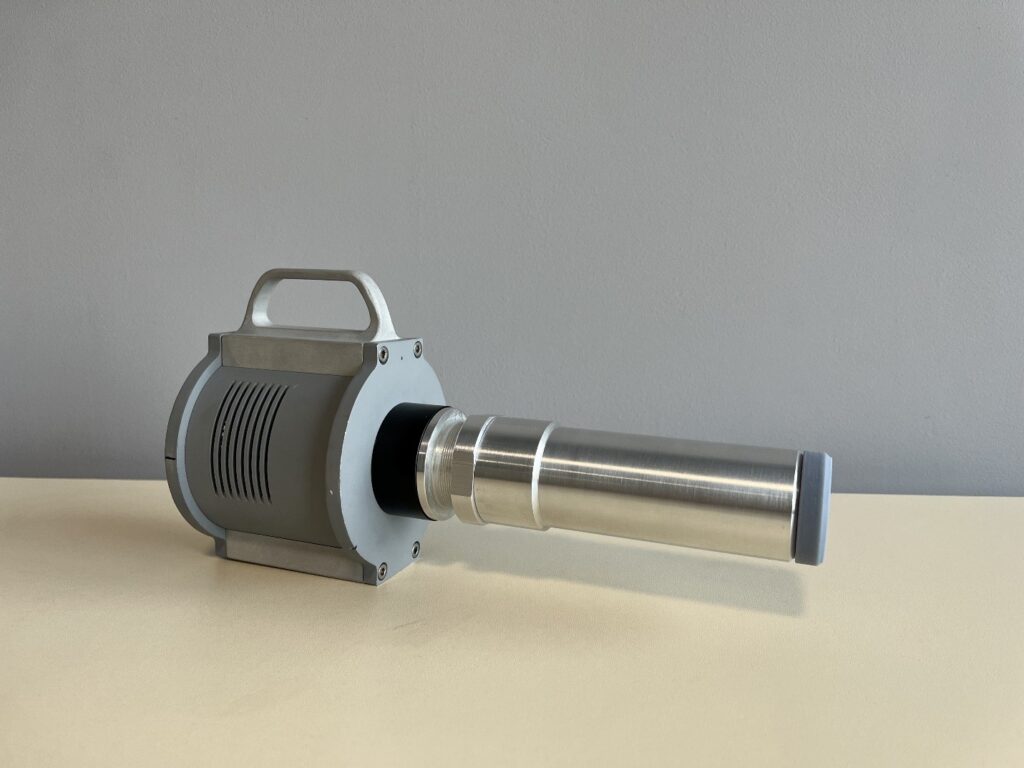
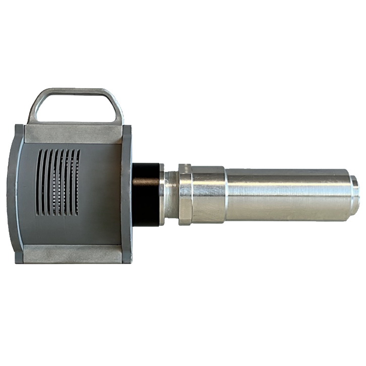
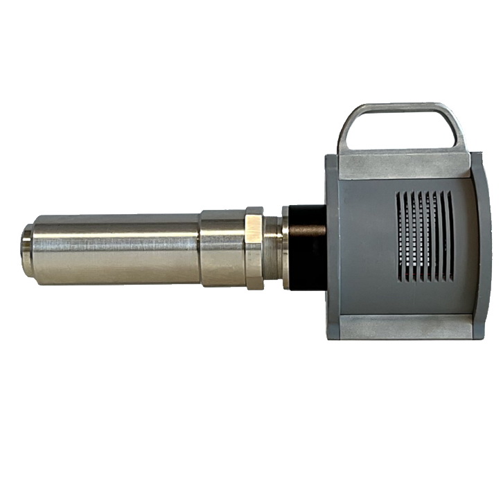
High-Resolution 2D X-Ray Detector «X-RAY EYE»
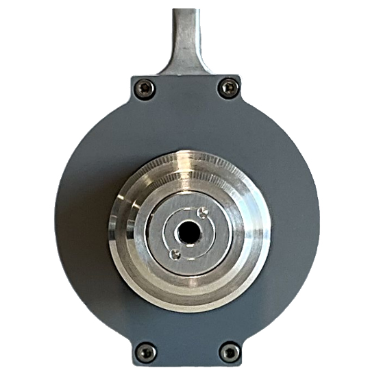
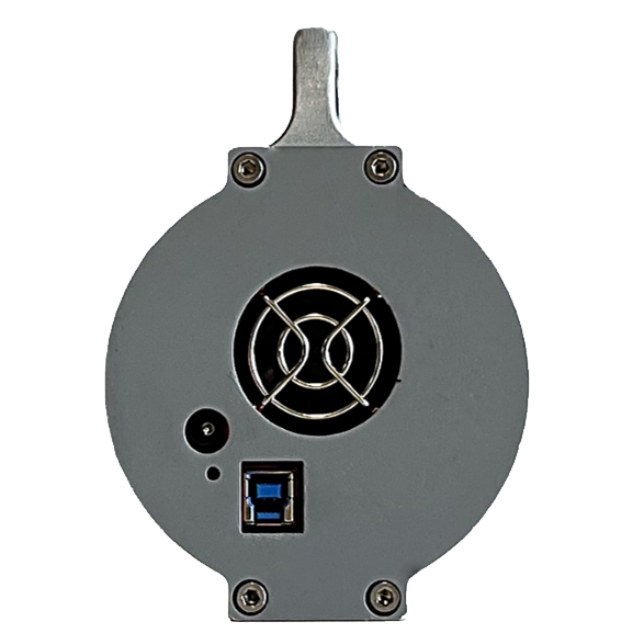
The X-ray compact high-resolution 2-D detector can be used both on laboratory X-ray sources and on sources of synchrotron radiation, incl. IV generation and XFEL (X-ray free electron laser). The detector’s main purpose is to visualize an X-ray image with a high (less than 10 µm) resolution. Applications include high-resolution imaging, X-ray microscopy, tomography, diffraction topography, diffractometry, and reflectometry. The device can be used for introscopy at stations: beam and instrumentation diagnostics, sample alignment, etc. The presence of hardware binning in combination with a wide exposure interval allows you to flexibly adjust the image acquisition mode. The detector is mounted with M4 or M6 screws on the bottom of the case, and small geometric dimensions (250.6mm x 96mm x 136mm) allow you to place the device in a limited space. The presence of active thermoelectric cooling makes it possible to significantly increase the signal-to-noise ratio, which makes it possible to use the detector in low-light conditions.
Specifications
| Imaging device | 20 MP Monochrome CMOS |
| Pixel size | 2.4×2.4 |
| Pixel array (horizontal x vertical) | 5496 x 3672 |
| Effective area (horizontal x vertical) | 4.7 x 3.2 mm |
| Readout noise | 1.6 – 3 e |
| Readout rate | 19 fps at full resolution |
| Full well capacity in electron | 15 000 |
| Digital output | 10- or 12-bit ADC |
| Resolution | 1 |
| Linearity error | 1 % or less |
| Exposure times | 0.032 – 2000 s |
| Binning | 2×2, 3×3, 4×4 |
| Cooling | Regulated two-stage thermoelectric cooling (TEC) |
| Geometry | 250.6x96x136 mm |
| Weight | 1.6 kg |
| Mount | 2 screw M6 or 3 screw M4 |
| PC connection | USB3.0/USB2.0 |
X-Ray Imaging
Conducting modern laboratory and synchrotron research is impossible to imagine without the use of detectors capable of visualizing X-rays. Such devices are used both at specialized synchrotron stations for high-resolution imaging, microscopy, tomography, topography, diffractometry, and can be used at non-specialized stations for introscopy, i.e., diagnostics and alignment of the X-ray beam, instruments, and sample. The design of such detectors was proposed and described in [1], and has not undergone fundamental changes since then. The detector in general form can be represented as a combination of a scintillator, an optical scheme (lenses or a set of lenses), and a digital CCD or CMOS matrix.
The parameters of the elements significantly impact the final characteristics of the detector. The main parameters of the detector, such as spatial resolution, speed, the field of view, etc., are determined by the application area and are set during the design and manufacture of the detector.
Detectors are manufactured using highly sensitive scintillation crystals with a thickness of tens to several hundred microns. The optical circuit collects the light generated by the scintillator and focuses it on the sensor. For lenses, an important requirement is a maximum transparency for the wavelength of the scintillator and the absence or minimization of aberrations.
The heart of the detector is the sensor, its parameters largely determine the characteristics of the device as a whole:
● ADC bit depth, widely used 8, 10, 12, 14, and 16-bit, this parameter determines how many gray levels the sensor can display.
● Imaging speed – a parameter showing how many images can be obtained per unit of time (fps). An increase in bit depth leads to a decrease in fps, therefore, for applications requiring high shooting speeds (fast processes or processes in biological tissues), you should choose cameras with 10 or even 8 bits, which can give a recording speed of up to 1000 fps.
● File size – in studies involving a large number of images (study of the dynamics of processes), large volumes of images are accumulated, the transfer and processing of which, due to their large volume, can be laborious and take a long time.
● Dynamic range – signal-to-noise ratio, in other words, the sensor’s ability to simultaneously display both very dark and very light details of an object.
● Gain – determines the number of photons per gray level. Often, different ADC bit depth allows you to vary the gain. This is important for visualization because different gain states have different levels of readout noise, making it important to choose the right bit depth and gain for low signal levels.
● Sensor pixel size is the physical size of one photosensitive element of the sensor. This parameter is important in experiments with low illumination, the signal/noise for a smaller pixel size will be less than for a pixel with large geometric dimensions.
● Effective pixel size and field of view. Using a lens system allows you to achieve the required level of magnification. In this case, the pixel may be significantly smaller than its physical dimensions. For example circuit magnification is 10x, the sensor pixel size is 10 µm, then the effective pixel size is 1 µm. This approach makes it possible to increase the spatial resolution of the detector. However, it should be understood that as the pixel size (effective size) decreases, the detector’s field of view also decreases.
A wide range of modern sensors provides a wide range of bit depth, fps, gain, and dynamic range. It is important to understand what works best for your application. Each imaging experiment is unique and works with different objects, light levels, and recording speeds, all of which influence the optimal choice of detector.
[1] – A. Koch, C. Raven, P. Spanne, A. Snigirev, J.Opt.Soc.Am.A., 1998, p. 1940 – 1951.